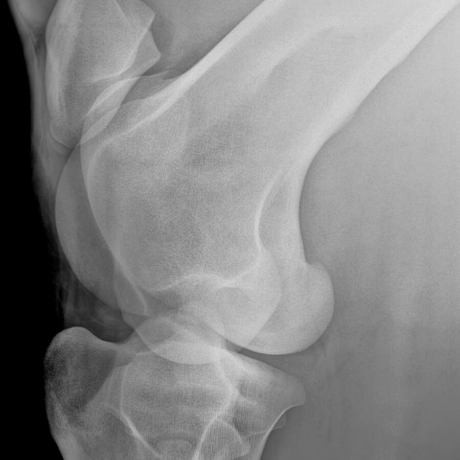
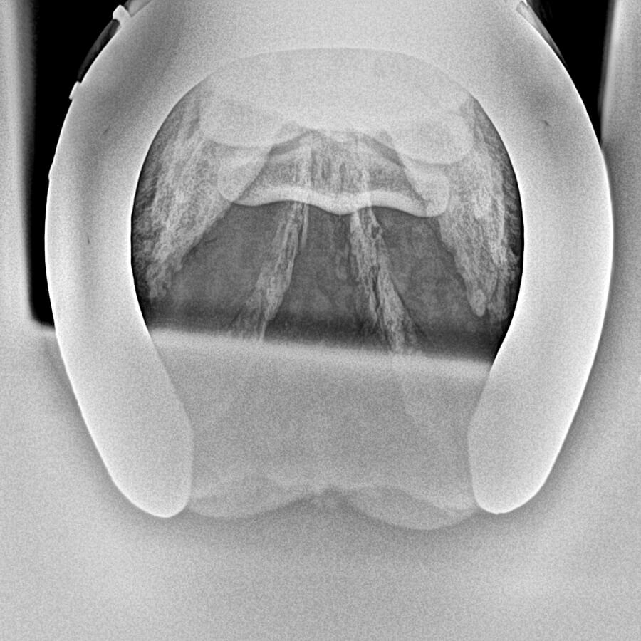
Surrey Hills Equine vets and nurses carry the latest and state-of-the-art portable wireless technology of diagnostic imaging tools, such as Digital X-ray systems, Ultrasound Scanners and Video Endoscopy directly to our clients’ premises. We provide state-of-the-art innovation to assist our clients’ horses with any clinical tool needed to aid an investigation at the same pace of a referral hospital, but directly at their premises, and hence provide them with the peace of mind of avoiding the need of transporting their horses away from their environment.
Additional diagnostic imaging may be advised by our vets to complement a clinical exam, may this be a lameness investigation, a pre-purchase examination (vetting), a dentistry or head-related problem, or an investigation of upper airways for respiratory conditions or for Strangles cases.
Furthermore, for those clients planning on buying a new horse, it is important to know that in case of pre-purchase examinations insurance companies may require to complement a 5-stage vetting with further diagnostic imaging to be able to obtain a comprehensive cover for veterinary fees and/or loss of use.
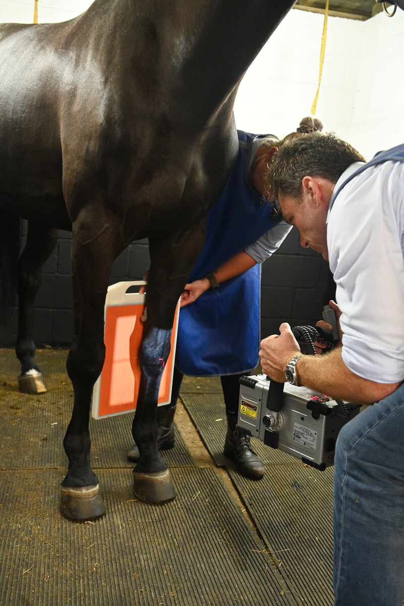

XRay
Xraying a horse can assist in making a more informed decision on a purchase. Xrays may be useful to assess for instance the health of navicular bones, or revealing the presence of Osteoarthritis (OA) which may in some cases lead to Degenerative Joint Disease (DJD).
When vetting younger horses, radiography may be most useful to assess hidden Osteochondritis Dissecans or Osteochondrosis (OCD), which is a common joint-related developmental abnormality. OCD commonly affects joint cartilage and can lead to the development of fragments (‘chips’) or subchondral bone cysts.
These are conditions that may increase the risk for future lameness and poor athletic performance in a horse, but also affect the ability of a buyer to successfully obtain veterinary insurance cover.
Xrays can also help a vet assessing angular limb deviation or deformities arisen in the developmental stage, such as crooked legs, pigeon toes, and general valgus, varus and windswept conditions.
Our vets will discuss any radiographic defect and advise on how these may affect the risk for use and resale, and the ability to purchase insurance.
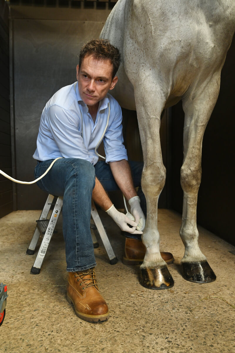
Ultrasound
Ultrasound scans can be used to assess size, shape and echogenicity (SSE) of soft tissues such as tendons or ligaments. ‘Tendinitis’ and ‘Desmitis’ are terms used to define inflammatory and traumatic events affecting tendons and ligaments. Ultrasound scans may discover ‘hypoechogenic’ or ‘hyperechogenic’ areas which may be associated to recent or older scarred lesions which may increase the likelihood of recurrence of lameness.
Ultrasonography may also allow to measure the cross-section area of equine tendons or ligaments which may carry an associated risk for lameness.
When vetting racehorses and racing ponies this may become particularly useful as it may allow to determine size, shape and echogenicity of Superficial Flexor Tendons (SDFT), Deep Digital Flexor Tendons (DDFT) and Suspensory Ligaments. Furthermore, a Suspensory Ligament Desmitis or Desmopathy is a frustrating condition that may present in both front and hind limbs of thoroughbreds, hunters, and sport horses, mostly jumpers and dressage horses.
This condition may cause a mild and intermittent lameness and may affect the origin, the body and the branches of a suspensory ligament. Whilst in the acute stages, a vet will be able to palpate a specific localised heat and swelling, but this will often not be present in any long-standing and chronic case. Ultrasound scans may therefore be useful to reveal rounded borders and enlargement of the suspensory, which may not necessarily be palpable and therefore may be missed during a clinical exam.
Highly, highly recommend this vet practice. Not only are they very professional and knowledgeable, but have the best kit in field medicine. Corrado is also kind and compassionate.
Call Now
Contact us now on
01306 249 944
or email info@surreyhillsequine.com.

